
“CT imaging contains an extraordinary amount of biological information, but historically we have reduced it to subjective impressions or crude categorical scores. Our AI models transform routine CT scans into precise quantitative biomarkers, allowing clinical trials to measure disease biology directly rather than relying solely on downstream functional surrogates.”
Dr Simon Walsh
Chief Scientific Officer
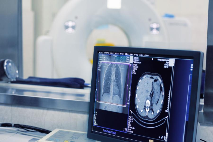
Our models are fully CT vendor-agnostic, validated across varying CT acquisition protocols, and require no respiratory gating or phantom calibration. Powered by full 3D volumetric deep convolutional neural networks, they deliver robust, reproducible quantification at scale. In peer-reviewed publications, our platform has demonstrated a 98.4% successful CT analysis rate.
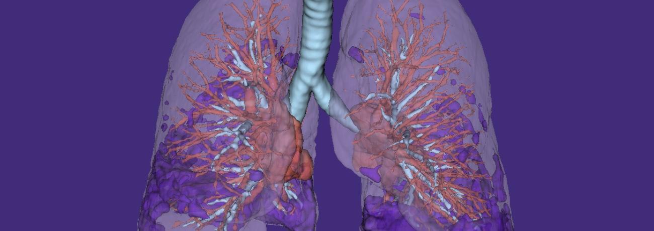
Each model isolates a biologically distinct lung compartment, enabling precise detection of compartment-level treatment effects.
- Fibr8™ quantifies lung fibrosis.
- Glass8™ quantifies inflammatory (ground glass) change.
- Air8™ quantifies airway volume.
- Vascul8™ quantifies pulmonary vascular volume.
- Lung8™ measures whole-lung and lobar volumes.
By measuring these compartments independently rather than relying on composite or indirect markers, Qureight enhances sensitivity to disease change, enables clearer interpretation of therapeutic impact, and provides compartment-specific mechanistic insight into drug effect
Beyond endpoint generation, Qureight provides real-time enrolment analytics
Imaging-derived biomarkers are analysed as patients are enrolled, allowing sponsors to monitor disease severity distributions and benchmark enrolled cohorts against predefined trial criteria and reference populations. This enables early detection of enrolment drift, supports proactive course correction during recruitment, and reduces late-stage trial failure risk by protecting cohort integrity before database lock.

-

Elucidation of a drug mechanism for a biotech partner
-

Analysis of combined imaging and proteomic data with a biopharma partner